Posterior Tibial Nerve Block
Welcome to the Posterior Tibial Nerve Block regional anesthesia page. Here we will cover indications, contraindications, materials and steps for you to master this procedure. Let’s begin with the overview below.
Overview
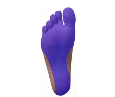
The posterior tibial nerve block is a safe, rapid, and effective method to provide analgesia to the calcaneus and plantar area of the foot. The sole of the foot can be a difficult area to anesthetize locally, and the posterior tibial nerve block requires fewer injections and a smaller volume of anesthetic compared to local anesthesia.[1] While this block can be performed using anatomic landmarks, the ultrasound-guided technique is associated with a higher success rate.[2]
Indications
Contraindications
There are few true contraindications to this block. They include:
- Infection (cellulitis) at the site of injection
- Allergy to local anesthetic being used
- Patient refusal
Contraindications
Some relative contraindications to consider:
- Patient needs a neural exam of the affected extremity.
- Anticoagulation use or bleeding disorder (pressure and tourniquet can be applied to the affected area if necessary).
- Prior neural lesion or active injury
Anatomy
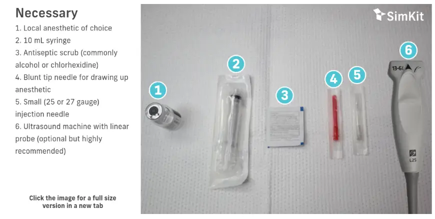
Materials
Technique
Ideally, blocks should be performed with aseptic technique using a skin antiseptic, sterile field, and sterile equipment if possible. Allow for appropriate dry time for your antiseptic. Block needles should be aspirated prior to every injection to avoid intravascular administration, and injection should be immediately stopped if the patient suddenly complains of pain or high pressure.
Landmark
Place patient in supine position with knee flexion and hip external rotation.
Palpate between the medial malleolus and Achilles tendon to find the injection site. It is described as 1-2 cm proximal to the medial malleolus and half way between the malleolus and Achilles tendon. Palpation of the posterior tibial pulse is also recommended the help guide injection away from the artery.
Clean the area with antiseptic solution.
Place a skin wheal of local anesthetic at the injection site.
Introduce the needle from medial to lateral with negative aspiration.
Inject 5mL of local anesthetic.
Ultrasound-Guided:
Place patient in supine position with knee flexion and hip external rotation.
Clean the area with antiseptic solution.
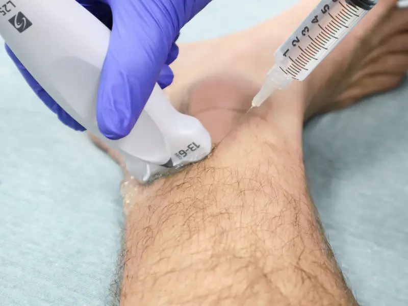
Position ultrasound probe in transverse position posterior and superior to medial malleolus (Figure 2).
Identify the tibial artery and vein. Identify the tibial nerve, posterior and lateral to the artery and vein (Figure 3).
Place a skin wheal of local anesthetic at the injection site.
Introduce needle at a 45 degree angle from anterior aspect in-plane to the probe (Figure 4).
After negative aspiration, inject 5mL of anesthetic superficial to the tibial nerve.
Summary & Bullet Points
- Posterior tibial nerve block is a safe and effective way to provide analgesia to the calcaneus and plantar surface of the foot.
- Very few contraindications exist (overlying infection, medication allergy).
- Ultrasound-guided technique is associated with a higher analgesia success rate.
Summary & Bullet Points
[1] Crystal CS, Blankenship RB. Local anesthetics and peripheral nerve blocks in the emergency department. Emerg Med Clin North Am. 2005; 23(2):477-502.
[2] Redborg, KE, Antonakakis JG, Beach ML,et al. Ultrasound improves the success rate of a tibial nerve block at the ankle. Reg Anesth Pain Med. 2009;34: 256-260.
[3] Clattenburg E, Herring A, Hagn C, et al. ED ultrasound-guided posterior tibial nerve blocks for calcaneal fracture analgesia. Am J Emerg Med. 2016;34:1183.e1-e3.
[4] WikEM contributors. Nerve Block: Foot. WikEM, The Global Emergency Medicine Wiki. Oct 12, 2021. Available at: https://wikem.org/wiki/Nerve_Block:_Foot. Accessed Oct 2022.
[5] Granger CJ, Cohen-Levy WB. Anatomy, Bony Pelvis and Lower Limb, Posterior Tibial Nerve. 2022 May 29. In: StatPearls [Internet]. Treasure Island (FL): StatPearls Publishing; 2022 Jan–. PMID: 31536230.